2021 / 2026
A software to measure radiation doses
New technology measures radiation doses without a dosimeter
Using cameras and computers to accurately measure radiation for doctors
People who work in operating theatres are exposed to a small amount of ionising radiation. In order to prevent health risks, dosimeters monitor individual radiation doses. However, these meters are not very accurate. SCK CEN, in partnership with six international partners, has developed a revolutionary technology, in order to calculate radiation doses more precisely using a camera and specialised software. This technology has great potential, according to the PODIUM project (Personal Online Dosimetry Using Computational Methods).
An interventional radiologist numbs the patient’s skin, pierces the groin artery and inserts a catheter into the blood vessel. The catheter has to be pushed to the area that he wishes to examine, before he injects a contrast agent. During this procedure, he takes x-rays of the blood vessels.
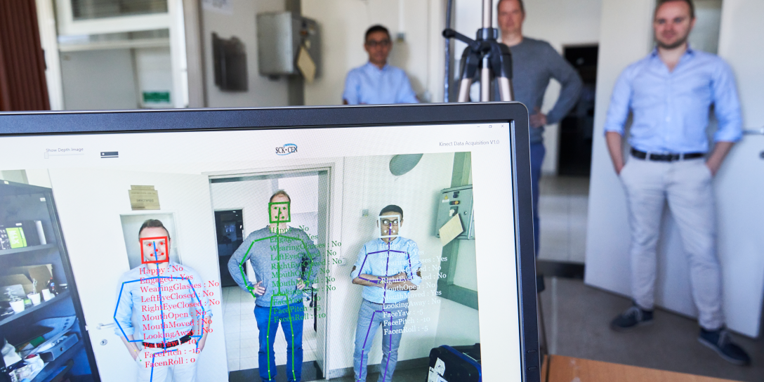
Most patients do not think about it, but their doctors are also exposed to ionising radiation. The radiation dose experienced by the doctor depends on his actions. For example, does he bend over the patient slightly when taking x-rays or does he hold his hands over them? This may increase the accumulated radiation dose. A conventional dosimeter measures the quantity of radiation inside the device. A camera and motion sensors make it possible to determine the radiation dose in each part of the body.
Gespecialiseerde software
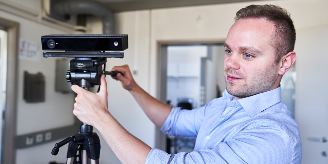
In partnership with six international partners, SCK CEN has developed a revolutionary technology in order to calculate the individual radiation dose experienced by a doctor. This is done by a camera and adapted software. The camera – together with motion sensors – closely monitors all the actions of the interventional radiologist and records every movement of his body. The software combines this data with the output from the x-ray machine being used, which enables it to calculate the radiation dose received. “The software takes into account the radiation field of the source and the distance between the radiologist and the patient”, explains SCK CEN scientist Pasquale Lombardo. “What energy level is the radiation? At which angle are certain parts of the body irradiated? It all makes a difference.”
Personalised dosimetry
The software used in the new technology was developed using simulations with dummies. This took place at St. James's Hospital in Ireland and the Malmö University Hospital in Sweden. The scientists placed anatomical torsos close to an x-ray machine. “We placed dosimeters inside the torsos in order to record exposure to radiation for the entire body,” explains Lombardo. “We collected the same measurements with and without personal protective equipment. For example, which dose would we measure in the different organs if the torso wore a lead apron? This is crucial information. We also used torsos of different sizes: male and female, big and small… This should make personalised dosimetry possible in the future.
Man of vrouw, groot of klein ... Onze nieuwe technologie biedt dosimetrie op maat.
More tests required
The technology seems to outperform the current physical dosimeters. Should we stop using them? “We need to walk before we can run”, says project coordinator Filip Vanhavere, with tempered expectations. “Physical dosimeters do indeed have limitations. For example, they only have one single measurement point for the whole body. As a result, they provide little information about the different organs. The measurement results are only available after a few weeks, with an uncertainty factor of 2 for measurement accuracy. Our software is able to overcome these limitations, but the technology is still in its infancy. It needs to be further developed and tested.”
The consortium partners plan to develop a prototype in 2021, before launching it in 2023 and marketing it in 2025. At the same time, they want to test it in other contexts, beyond interventional radiology and cardiology. Last year, the scientists already installed a test configuration in workplaces with neutrons.
Innovative approach
The new technology is part of the regulatory principle known as ALARA (“As Low As Reasonably Achievable”). This principle stipulates that a radiation dose must always be kept as low as possible. Other scientists from all over the world have also used cameras with motion sensors to monitor the ALARA principle. As soon as a person comes too close to a radiation source, a warning appears. But the PODIUM project is taking things further.
Vanhavere: “We use cameras for personalised dosimetry. This approach is unique and an international innovation. In addition, computers are becoming faster and faster. This development is important, as we calculate the organ doses based on “Monte-Carlo” simulations. A Monte-Carlo simulation is a computer-controlled technique, which simulates a physical process not once but many times. In our research, this physical process involves emitting radiation particles.”
“In a Monte-Carlo simulation, we release millions of radiation particles from the x-ray machine”, explains Lombardo. “Each radiation particle has a different energy or flies in a different direction. We follow all of them on their different paths and calculate the radiation dose that they ultimately deposit in the different organs. Every time the doctor moves, we repeat the same simulation. This requires powerful and therefore fast computers, but it also produces very accurate data. The more powerful computers become and as the quality of images from the camera continue to improve, the more we can further refine our technology.”

Seven partners pulling together
Seven partners joined forces to make the innovative PODIUM project a success: SCK CEN (Belgium), Universitat Politècnica de Cataluyna - UPC (Spain), Helmholtz Zentrum Munich (Germany), Lund University (Sweden), Public Health England (Great Britain), Greek Atomic Energy Commission (Greece) and St. James's Hospital Ireland (Ireland). “Monitoring individual radiation doses is crucial for effective radiation protection. Our goal is to continue our research and improve our current measuring techniques”, says Filip Vanhavere.
The PODIUM project is funded by the Euratom 2014- 2018 Research and Training Programme. It forms part of the H2020 CONCERT project, a European programme integrating European and national radiation protection research.
We used cameras for personalised dosimetry. This approach is unique and an international innovation.
The fourth industrial revolution merges the physical, digital and biological worlds. Also the world of radiation protection basic science is improved with advanced technological developments, artificial intelligence, machine learning, and big data. They are used for increased protection of humans and their environment in the context of medical applications, advanced impact studies, space missions… Innovative and rigorous, an indispensable link in a multidisciplinary network.
— Hildegarde Vandenhove (Environment, Health and Safety)